 Home
Home- About
- Resources
- Community
- Community
- PVED Forum
- Calendar & Events
- Our Blog
- DE Medical Info
- DE Legal Info
- DE Insurance Resources
- DE Insurance Resources
- Insurance Advocacy
- DE Articles
- DE Surrogacy
- Embryo Donation
- Embryo Donation
- Embryo Donation Programs
- Donate Your Embryos
- PVED Embryo Donation Program
- DE Emotional Health
- DE Emotional Health
- Building Family Through Embryo Donation
- Talking To Kids About Donor Conception
- Why Should I See A Therapist?
- Positive Family Building Language
- Getting Ready, Letting Go
- Children & Understanding Family Building
- Recommended Reading List
- Contact Gamete Donors
- Faculty Biography
- Open Identity
- Children & Conception Talk
- DE IVF Abroad
- Donor Egg Banks
- Donor Egg Banks
- Comparing Egg Banks
- Frozen Oocytes
- Egg Banking FAQ
- Egg to Baby
- FAQ
 Donate
- Contact
 Advertise
 Site Map

 “PVED provided the information, help, and support I needed to navigate the building of our family.”
--Cristin
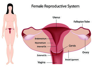 A Hysterosalpingogram (HSG) is an X-ray test that looks at the inside of the uterus and fallopian tubes and the area around them. It often is done for women who are having a hard time becoming pregnant.
When a woman undergoes an HSG dye which is known as contrast material is then pushed through a thin little tube that’s introduced into the vagina, through the cervix and then into the uterus. Since the uterus and our fallopian tubes are connected or hooked together if you will, the dye flows through the fallopian tubes. While this is happening the technician is taking pictures using a medical imaging technique called fluoroscopy which is a steady beam of X-ray. When the dye passes through the uterus and the fallopian tubes the pictures (images) can show issues within the uterus or the fallopian tubes. For instance if the uterus or fallopian tubes have been injured or there is some sort of an abnormality that would prevent an egg from dropping through the fallopian into the uterus, or in our case something that would prevent implantation of an embryo that’s been placed back into the uterus during an embryo transfer.
A Hysteroscopy is a great way for your doctor to look at the lining of your uterus. Your doctor will use a thin little tool called a hysteroscope. The tip of the hysteroscope is put into your vagina and your doctor will slow and gently move this tool through the cervix into the uterus. The hysteroscope has a light and camera hooked to it so your doctor can see the lining (endometrium) on a video screen. Doctors may want to perform a hysteroscopy on a patient to determine why they may be experiencing abnormal bleeding in their uterus, or loo examine the uterus to look to see if there’s anything in the uterus that may not belong that could be preventing implantation of an embryo within the uterus.
A hysteroscopy can be used to remove growths in the uterus, such as fibroids or polyps. Your doctor may take a tissue biopsy of your uterine lining and examine that tissue under a microscope for problems.
Laparoscopy is a surgery that uses a thin, lighted tube put through a small cut (incision) in the belly to look at the abdominal organs camera or the female pelvic organs camera. Laparoscopy is used to find problems such as cysts, adhesions, fibroids camera, and infection. Tissue samples can be taken for biopsy through the tube (laparoscope).
This procedure is done to look for and remove abnormal growths like tumors or polyps in the uterus, belly or the pelvis. During this procedure doctors can check for and treat things like ecoptic (tubal) pregnancies, endometriosis, or pelvic inflammatory disease (PID)
When a woman is trying to get pregnant things like cysts, fibroids, infections, or adhesions can be treated as well.


